[FILM-Users 00615] FW: CRL Seminar | Wednesday 24th February, 3pm | Neil Chalmers SR | Lidia Sonakowska | Visualising crustacea digestive tract using freshwater shrimp as a model organism
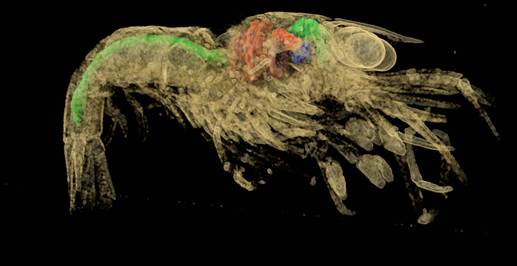
FYI: see below From: Tomasz Goral [mailto:T.Goral@nhm.ac.uk] Sent: 22 February 2016 14:08 To: Keller, Debora Subject: FW: CRL Seminar | Wednesday 24th February, 3pm | Neil Chalmers SR | Lidia Sonakowska | Visualising crustacea digestive tract using freshwater shrimp as a model organism Dear Deborah, This Wednesday we've got an interesting seminar from our Synthesys visitor showing different visualisation approaches for studying crustacean development. I thought it might be interesting for some of your FILM users. Would you mind circulating it as I don't seem to have a FILM users address? Many thanks, Tomasz Dr Tomasz Goral Electron Microscopist Imaging and Analysis Centre The Natural History Museum London SW7 5BD Tel. 02079425403 http://www.nhm.ac.uk/research-curation/science-facilities/analytical-imaging... From: Tomasz Goral Sent: 16 February 2016 16:33 To: Science-Group Cc: Synthesys Alerts Subject: CRL Seminar | Wednesday 24th February, 3pm | Neil Chalmers SR | Lidia Sonakowska | Visualising crustacea digestive tract using freshwater shrimp as a model organism Visualising crustacea digestive tract using freshwater shrimp as a model organism. Lidia Sonakowska Department of Animal Histology and Embryology, University of Silesia, Bankowa 9, 40-007 Katowice, Poland Neil Chalmers Seminar Room, 3pm, 24th February [Wy?wietlam 12.png] Arthropods are the most diverse and common animals that live all over the world. Crustacea, which belong to that group show an incredible diversity of morphology, size, and habitats attesting their evolutionary success. Although they are the most abundant animals which mainly live in aquatic ecosystems, the knowledge about their morphology and development is rather fragmentary and there is still a lot of information which needs to be clarified. Moreover, most studies of anatomy and morphology of decapods are based on classical methods. The lack of general information on development and histology of crustacea digestive tract encouraged me to start this study. In my talk I will present a comparison of novel vs classical histological and embriological techniques employing high resolution microscopy and micro-CT scanning for 3D visualisations of the structure and ultrastructure of digestive system of freshwater shrimp Neocaridina heteropoda (Crustacea, Malacostraca, Decapoda) which can serve as great example for comparative study for other crustaceans. Futhuremore, I will show the importance of regeneration and degeneration processes which occur in midgut epithelium in development, proper functioning and homeostasis maintenance of the investigated organ. Dr Tomasz Goral Electron Microscopist Imaging and Analysis Centre The Natural History Museum London SW7 5BD Tel. 02079425403 http://www.nhm.ac.uk/research-curation/science-facilities/analytical-imaging...
participants (1)
-
Keller, Debora