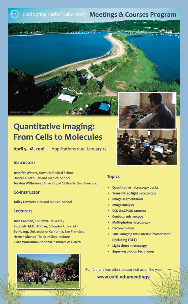
Dear users, See below I wanted to let you know about a quantitative microscopy course that Torsten Wittmann (UCSF), Hunter Elliott (Harvard), Talley Lambert (Harvard) and I are organizing at Cold Spring Harbor Laboratory, called "Quantitative Imaging: From Cells to Molecules". We will be teaching the course for the 5th time April 5-18, 2016, with applications due January 15, 2016. Our course has received excellent reviews from the students, and we feel it has been a great success. We accept only 16 students, and we have excellent support from vendors in the form of equipment loans - so each student gets lots of hands-on time with state-of-the-art microscopes during the labs. We also emphasize quantitation throughout the course, both in lectures and in daily quantitative lab exercises. The course is supported by a grant from NCI, so CSHL is able to offer financial aid to many students to help cover tuition costs. Course Description: Combining careful image acquisition with computational analysis allows us to extract quantitative data from light microscopy images that is far more informative and reliable than what can be seen by eye. This course will focus on advanced quantitative fluorescence microscopy techniques used for imaging a range of biological specimens, from cells to single molecules. The course is designed for cell and molecular biologists with little or no microscopy experience who wish to begin utilizing microscopy in their own research. Students will gain a theoretical understanding of, and hands-on experience with, state-of-the-art equipment used in quantitative fluorescence microscopy, including: wide-field fluorescence microscopy, laser scanning and spinning disk confocal microscopy, total internal fluorescence microscopy (TIRF), super-resolution methods (structured illumination, STED, STORM and PALM) and digital image processing and analysis. Students will learn how to design and implement a wide range of imaging experiments using these techniques. Students will also learn fundamental image processing, segmentation and analysis techniques using a variety of commercial and open source software packages. Students will use these image acquisition and analysis techniques to address specific quantitative questions and then discuss the results as a group, learning to troubleshoot the common problems that occur in the course of a quantitative imaging experiment. Among the lectures presented are: quantitative microscopy basics, transmitted light microscopy, image segmentation, image analysis, CCD & sCMOS cameras, confocal microscopy, multi-photon microscopy, deconvolution, TIRF, imaging ratio-metric "biosensors" (including FRET), light sheet microscopy and super-resolution techniques. Students will also learn guidelines for choosing fluorescent proteins, and work with live samples requiring environmental control. Additional Lecturers for 2016: Julie Canman, Columbia University Elizabeth M.C. Hillman, Columbia University Bo Huang, University of California, San Francisco Nathan Shaner, The Scintillon Institute Clare Waterman, National Institutes of Health More info and online application: http://meetings.cshl.edu/courses.aspx?course=c-qicm&year=16 Best, J Jennifer C. Waters, PhD :: Director of Microscopy Harvard Medical School :: Dept of Cell Biology :: 240 Longwood Ave :: Boston, MA 02115 (617) 432-3542 :: jennifer_waters@hms.harvard.edu<mailto:jennifer_waters@hms.harvard.edu>